
Medical Services
Cardiological Consultation
Consulting means understanding. First we listen to you when you describe your situation and your problems. Then we ask specifically for connections and accompanying circumstances and take into account preliminary findings that are already available. Finally, we recommend which steps are sensible for you to take in order to achieve a result.
General Cardiology
General cardiology is important for every patient. It deals with frequently occurring disorders and diseases of the heart such as high blood pressure, rhythm disturbances, cardiac insufficiency. In addition to routine diagnostics and therapy, aspects of lifestyle and correct behaviour are also of great importance.
Cardiological Examination with Evaluation of the Cardiac Output
The cardiac complete examination with assessment of the heart performance (cardiac health check-up) consists of a medical history, a physical examination including blood pressure measurement, examination of the urine for blood, protein and glucose (sugar) as well as the determination of cholesterol and glucose in the blood. At the end of the examination, a final consultation takes place in which the doctor informs the patient about the findings and, if necessary, makes recommendations for further examinations. The cardiac complete examination is used for the early detection of frequently occurring diseases that can be effectively treated and whose early stage can be determined by diagnostic measures. These include cardiovascular and kidney diseases, especially hypertension, and diabetes mellitus. As these diseases increase from the age of about 35 years, a basic check-up is recommended for all women and men from the age of 36 (from 35 years) every two to five years.
Cardiovascular-Specific Applications and Specialized Consultation Hours
Serious heart diseases such as blockages of the coronary arteries, changes in the heart valves, heart failure and complex rhythm disturbances require special knowledge and examination methods. In our practice, the entire spectrum is available, if necessary also in cooperation with the University Hospital of Basel.
Prevention
Preventive measures play a special role, especially in the case of heart disease in the family or heavy work or family stress. After a risk profile has been drawn up (see also Heart Check), the importance of the existing risk factors (e.g. stress, job, family, sport, metabolism, blood pressure, etc.) is assessed and a prevention programme is drawn up.
Cardiac Risk Check-Up
In addition to risk analysis (see also prevention), the heart check includes examination of the heart with regard to structural changes, performance and resilience. Ergometry, spiroergometry, cardiac echo and special laboratory tests are used as required. Whether you are healthy or ill, you will find out exactly what stress your heart can be expected to endure in everyday life, while travelling and doing sports, and where the limits lie.
Training Management and Planning
Healthy, but above all sick people and athletes should train their heart under medical supervision if possible. A step-by-step plan individually tailored to the personal situation of an illness or training goal allows for appropriate and healthy performance development with rapid success without overloading.
Follow-up Care
Careful aftercare, whether after a heart attack, after cardiac catheterization with stent implantation, after pacemaker implantation or after any other form of heart surgery, serves to ensure the success of treatment. Regular routine examinations, even if the patient's subjective well-being is affected, are therefore also important under the aspect of prevention before the respective heart disease progresses.
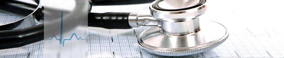
Electrocardiogram (ECG)
The electrocardiogram, ECG for short, is one of the basic examinations in medicine. The fine electrical currents and impulses of the heart muscle are recorded. These provide basic information about the heart rhythm, the excitation conduction and possible heart damage.
Ergometry (Stress ECG)
In ergometry, the ECG is recorded during physical exercise (stationary bicycle). This serves to detect circulatory disturbances in the coronary vessels, increases in blood pressure or cardiac arrhythmia that are not manifest at rest.
Long-term (Holter) ECG and Event Recorder
To evaluate cardiac arrhythmias in everyday life, especially if they occur irregularly (heart stumbling) or even unnoticed, a long-term ECG is recorded over 24-72 hours. A small recorder, which is attached to the belt or under clothing, stores the ECG, which is continuously derived via three adhesive electrodes, and is then evaluated by a computer.
An event recorder can be useful for rarely occurring rhythm disturbances. This is a mobile memory device the size of a cheque card, which is held on your chest in case of a subjectively felt heart stumble, records the heart rhythm and, if necessary, transmits it to our practice via a telephone connection.
Long-Term Blood Pressure Measurement
In contrast to the occasional blood pressure measurements at the doctor's office or the blood pressure self-measurements at home, the long-term blood pressure measurement by means of devices also allows an assessment of the blood pressure behaviour in everyday situations and during sleep. This information is crucial for careful individual blood pressure adjustment.
For this purpose, a blood pressure cuff is placed on your upper arm, which is connected to a small recorder that you wear on your body. Blood pressure measurements are then taken at regular intervals and can also be triggered by you.
Echocardiography With High-End 4D cSound Technology
Echocardiography refers to the examination of the heart by ultrasound and, in addition to the resting ECG, is the basic diagnosis for almost all cardiological questions. The ultrasound examination makes it possible to show the heart walls and heart valves. In addition, the blood flow in the area of the heart can also be depicted in colour (colour Doppler, duplex). The measurements that can be carried out in this way allow the reliable assessment of diseases of the heart muscle (thickening, weakness, heart attack), the heart valves (narrowing, leakage) and of heart defects. We perform the examinations using the latest GE E95 ultrasound system with state-of-the-art 4-dimensional cSound technology, which allows for highly precise, state-of-the-art non-invasive diagnostics.
Stress Echocardiography
Stress echocardiography is the combination of echocardiography and ergometry, i.e. heart ultrasound during physical stress. This allows stress-related circulatory disorders of the heart muscle to be recorded more sensitively than with ergometry alone. Thus, stress echocardiography can often make a possible hercocatheter examination unnecessary or necessary. Stress echocardiography also provides important information for special problems, for diseases of the heart valves (e.g. aortic valve stenosis) or the heart muscle (thickening), which can be decisive for therapy. The heart is stressed by the administration of a heart stimulating drug (dobutamine) via a vein. The patient lies calmly on the examination couch and does not have to pedal on a bicycle, so that the ultrasound images of the heart are not disturbed by physical movements and can therefore be derived in optimal quality.
Transesophageal Echocardiography (TEE)
Certain structures of the heart cannot be visualized sufficiently well by means of external ultrasound (transthoracic echocardiography). In these cases, a special form of ultrasound examination of the heart may be necessary, namely transesophageal echocardiography from the esophagus. For this purpose, a slim ultrasound probe is inserted into the esophagus via the throat (similar to a gastroscopy), and the heart is examined from close proximity from the inside. To make the examination as comfortable as possible for you as a patient, a local anaesthetic is applied to the throat beforehand and, if necessary, a sedative is administered. Swallow echoes are normally performed by us in cooperation with the University Hospital of Basel.
Ergospirometry (a.k.a. Spiroergometry)
Ergospirometry is an exercise ECG (ergometry), in which the lung function (spirometry) is additionally determined via a breathing mask and the concentrations of oxygen (O2) and carbon dioxide (CO2) in the air breathed are determined for each breath. This serves to provide precise information on functional disorders of the heart, lungs and metabolism under stress. In the case of pathological limitations in performance (symptom breathlessness), spiroergometric analyses provide important information about causes in the cardiac (heart), pulmonary (lung) or metabolic (metabolism) areas. Therefore cardiologists, pulmonologists, diabetologists, but also occupational and rehabilitation physicians use ergospirometry. In training planning and sports medicine, ergospirometry is now the basis of professional training consultation and training planning (much better statement than the lactate test). If indicated, we perform ergospirometry by referral to the university hospital and incorporate the results into our overall diagnosis.
Cardiac Pacemaker - Implantation, Check-Up and Programming
If the heart beats too slowly or if several heart actions fail, this can lead to dizziness, weakness or loss of consciousness and even to a life-threatening circulatory arrest. By implanting a pacemaker, the heart rhythm can be restored.
The pacemaker unit is the size of a matchbook and is inserted under the skin below the collarbone under local anaesthetic. From there, thin probes are inserted through a vein into the heart and the pacemaker stimulates the heart at the required rate. We usually perform the procedure by referral to the University Hospital of Basel with a short in-patient stay at the hospital.
After the implantation, a regular check of the pacemaker system is necessary, usually at 6-month intervals. We check the technical functionality and the battery condition. In the course of time, it may become necessary to adapt the programming of the pacemaker to new requirements due to changes in life circumstances or changes in the heart. These adaptations are then carried out again by referral to the University Hospital of Basel.
Cardiac Resynchronisation Therapy (CRT), 3-Chamber or conduction system pacing
The normal pacemaker, which is implanted when the heartbeat is too slow or absent, has a probe in the right ventricle, sometimes another one in the right atrium, to deliver electrical pulses. In case of a higher degree of cardiac insufficiency and the presence of a left bundle branch block, a resynchronization pacemaker can be implanted, which has an additional probe near the left ventricle. This allows the coordination of the left and right ventricle to be restored (synchronization), resulting in improved performance. Resynchronization pacemakers are often combined with implantable defibrillators in one unit. The implantation of the resynchronization aggregates is performed by referral to the University Hospital of Basel in its cardiac catheter laboratory. Like the implantation of an ordinary pacemaker, it is performed under local anaesthesia.
Implantable Cardioverter-Defibrillator (ICD)
Ventricular fibrillation or ventricular tachycardia are particularly dangerous and sometimes fatal cardiac arrhythmias. Implantable defibrillators have been developed to stop these arrhythmias and bring the heart back into rhythm. Depending on requirements, this can be done by a special type of electrical heart stimulation or by delivering a current pulse (defibrillation). These aggregates are implanted in the chest area like pacemakers.
The control by an experienced heart specialist should normally take place at 3-month intervals. In addition to the technical functionality, we also check whether the unit has recorded any unnoticed cardiac arrhythmia in the meantime. This information is important for the planning of medication therapy as well as for the programming of the devices.
Intracardiac Catheter
An essential component of modern cardiological examination methods is the heart catheter examination, which can include diagnostics and therapy in one examination procedure. Nowadays, the so-called minimally invasive heart catheter technique via the artery at the wrist is of particular importance. This gentle procedure is particularly suitable for outpatient cardiac catheter examinations. Cardiac catheter examinations are performed by referral to the University Hospital of Basel. Your cardiologist, who is already familiar with the procedure from our practice, will personally accompany you through the procedure and continue to look after you afterwards.
In the diagnostic part of the examination the coronary vessels are probed with a thin catheter and filled with X-ray contrast medium. The whole procedure is recorded as a video film via an X-ray machine. The cardiac catheter laboratory of the University Hospital of Basel is equipped with the latest generation of X-ray equipment, which, thanks to digital image management, makes it possible to keep the patient's exposure to radiation to a minimum.
If the evaluation of these coronary angiography films does not yet allow a reliable assessment of displayed coronary artery stenoses, two additional diagnostic techniques may be used if necessary: Firstly, a small ultrasound probe can be inserted into the coronary vessel (intravascular ultrasound, IVUS) and thus the exact fine structure of the coronary vessel and the existing constrictions can be examined. Or a special small pressure sensor can be positioned in the coronary vessel in front of and behind the constriction, and the significance of the constriction can be precisely determined by the course of the pressure measurements. By using this technique in a sensible way, unnecessary stent implantations can be avoided.
If the diagnostic part of the cardiac catheter examination has revealed a significant narrowing, it is in most cases advisable to treat this stenosis directly in the same session. Here too, it is important to choose the most suitable method for you personally from the multitude of possible methods. In addition to the usual routine procedures such as balloon dilatation (coated and uncoated) and stent implantation (coated and uncoated), so-called rotablation is also available. In this procedure, a very small, diamond-studded milling head is inserted into the coronary vessel, which then opens the constriction at high speed. This technique is mainly used for hard calcified constrictions that cannot be dilated with the usual balloons.
Electrophysiology and Catheter Ablation
In electrophysiology, rhythm disturbances are treated by catheter ablation. The aim of catheter ablation is the permanent elimination of cardiac arrhythmias by obliterating the heart muscle tissue responsible for them.
By using high-frequency current (frequency 500 kHz, power 10-60 watts, depending on the application also with cooled ablation technology), the surrounding heart muscle tissue is heated to approx. 55-65 degrees Celsius, resulting in punctual sclerotherapy with formation of a circumscribed scar (approx. 2-3 mm diameter).
In special situations, ablation with refrigeration technology (cryoablation with catheter or balloon with up to -80 degrees Celsius) can be used.
Catheter ablation can be successfully used in various arrhythmias, especially in WPW syndrome (excess conduction between the atrium and the main chamber), in AV node tachycardia (AV node reentry tachycardia), in typical atrial flutter of the common type (reentry, i.e. circular excitation around the tricuspid valve), in atrial tachycardia (tachycardia of the atrium), in atypical atrial flutter (high-frequency atrial (reentry) tachycardia), in ventricular extrasystoles (VES), in ventricular tachycardia (ventricular tachycardia, tachycardia from the left or right main chamber), in atrial fibrillation and in complex arrhythmias after heart surgery (circular excitation (reentry) around postoperative scars).
We perform catheter ablations by referral to the University Hospital of Basel.
Second Opinion and Expertise
A medical second opinion is the second, independent assessment of an initial medical finding by a second doctor. The second opinion can refer to a disease or a treatment measure. Obtaining a second medical opinion can help to avoid a false diagnosis or simply make the patient feel more confident by removing doubts about a finding. In particular, before taking serious therapeutic measures, as is the case with cardiological problems, it is worth asking a second doctor for advice, for example when it is necessary to choose from various therapeutic options. However, even in the case of surgical operations that require a longer stay in hospital or treatment with drugs that have many side effects, it can be useful to have two doctors assess their necessity. Due to the extensive experience of the Cardiology Center, we offer reliable and precise second opinions and expertises in the entire cardiac field.